Sample Questions
1 of 20
Q
Which of the following is the most correct statement based upon imaging:
2 of 20
Q
A4C cine image is shown below
Findings are consistent with which of the following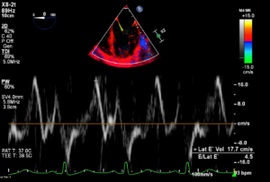
3 of 20
Q
PLAX M Mode is shown below. Cursor line is placed through the aortic valve. Findings are consistent with
4 of 20
Q
Severe AR is indicated by which of the following:
5 of 20
Q
Calculation of Effective Regurgitant Orifice Area (EROA) by the PISA method is an example of
6 of 20
Q
Aortic regurgitant volume calculation by flow convergence (PISA method) uses which of the following equations:
7 of 20
Q
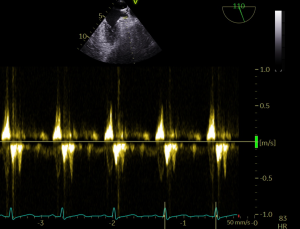
Suprasternal image is shown below. Combined colour Doppler and M mode of flow in the descending aorta is shown. Findings are consistent with
8 of 20
Q
Aortic valve dimensionless index is defined by which of the following
9 of 20
Q
Incorrect measurement of LVOT diameter resulting in underestimated LVOT diameter will have which effect on subsequent estimation of AS severity
10 of 20
Q
Estimation of mitral regurgitation severity is affected by severe aortic stenosis in which way
11 of 20
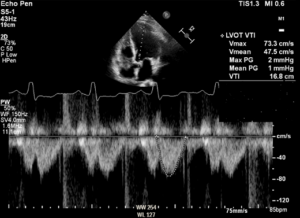
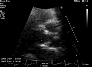
Q
Echo data are shown below. If LVEF is 35% and BSA (body surface area) is 1.8m², findings are consistent with which of the following: 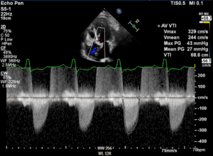
12 of 20
Q
Which mitral valve scallops are visible in A2C
13 of 20
Q
Waveform from CW Doppler interrogation of mitral valve inflow is shown below. Findings are consistent with which of the following
14 of 20
Q
HCM -SCD (Hypertrophic Cardiomyopathy – Sudden Cardiac Death) risk scores are used to calculate SCD risk in HCM patients. Which of the following echo parameters are used as part of the scoring system:
15 of 20
Q
Restrictive cardiomyopathy and Constrictive pericarditis may be distinguished based on the following:
16 of 20
Q
PW Doppler interrogation of mitral valve inflow is shown below. E/A ratio is consistent with which grade(s) of diastolic dysfunction:
17 of 20
Q
Mitral inflow and mitral TDI data are shown below 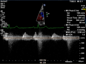
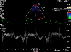
Findings are suggestive of:
18 of 20
Q
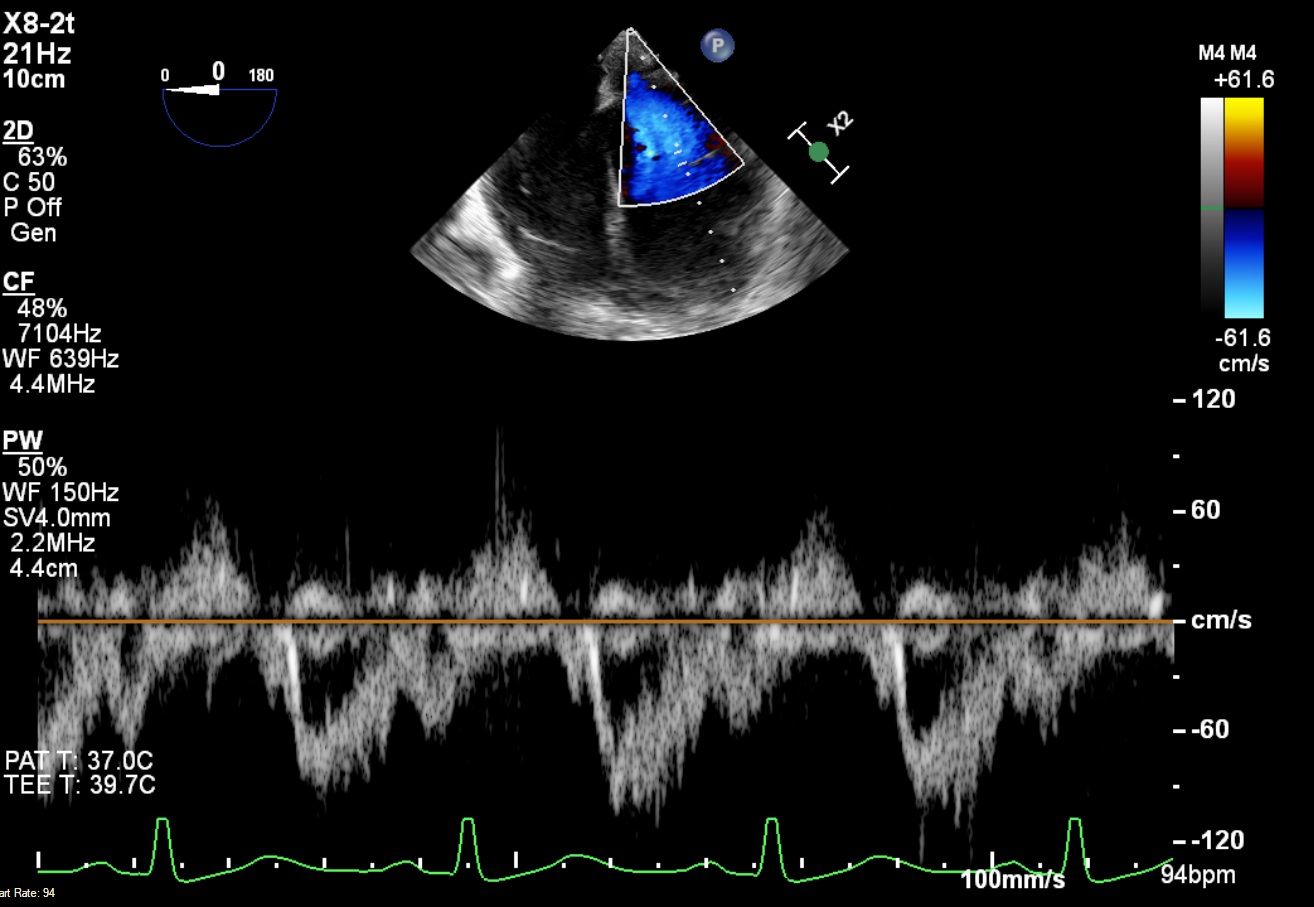
PW Doppler interrogation of pulmonary venous flow is shown below. Assuming duration of mitral inflow A wave (Adur) is 105msec, findings are suggestive of
19 of 20
Q
Echo sweep speed for assessment of respiratory variation in mitral and tricuspid valve inflow is best described by
20 of 20
Q
Aortic valve morphology in PSAX aortic valve level is shown below
Findings are consistent with which of the following